What are the symptoms of phyllodes tumors?
Phyllodes tumors, also known as cystosarcoma phyllodes, are rare breast tumors that can vary significantly in their presentation. The symptoms of phyllodes tumors include:
1. Breast Mass:
- Palpable Lump: The most common symptom is a noticeable, often large, lump in the breast. This lump is usually well-defined, smooth, and can be mobile under the skin.
- Rapid Growth: Unlike most breast tumors, phyllodes tumors can grow quickly and may become quite large, sometimes reaching several centimeters in diameter.
2. Changes in Breast Appearance:
- Size Increase: The affected breast may appear larger or asymmetrical compared to the other breast.
- Skin Changes: In some cases, the skin over the tumor may become stretched, or there may be dimpling or redness, although this is less common.
3. Pain or Discomfort:
- Variable Pain: Some patients may experience tenderness or discomfort, but many phyllodes tumors are painless. The level of pain or discomfort can vary depending on the size and location of the tumor.
4. Nipple Discharge:
- Uncommon: While less common, some patients might notice a discharge from the nipple, which is typically not the primary symptom.
5. Enlarged Lymph Nodes:
- Secondary: Although phyllodes tumors generally do not metastasize to lymph nodes, in rare cases, regional lymph nodes may be affected if the tumor is particularly aggressive.
6. Systemic Symptoms:
- Rare: Systemic symptoms such as weight loss or fatigue are rare and usually occur in more advanced cases where the tumor has spread beyond the breast.
Summary:
Phyllodes tumors are characterized by a rapidly growing breast mass that is typically well-defined and mobile. They can cause changes in breast appearance and, in some cases, discomfort. The presence of a large, growing lump is the most common symptom, and any significant changes in the breast should be evaluated by a healthcare professional. Early diagnosis and treatment are crucial for managing phyllodes tumors effectively.
What are the causes of phyllodes tumors?
The exact cause of phyllodes tumors is not well understood, but several factors have been associated with their development:
1. Genetic Factors:
- Genetic Mutations: There may be genetic mutations or alterations involved in the development of phyllodes tumors, although specific genes have not been definitively identified.
- Familial Syndromes: In some cases, phyllodes tumors may occur in individuals with certain familial cancer syndromes, such as Li-Fraumeni syndrome, although this is rare.
2. Hormonal Factors:
- Hormone Sensitivity: Phyllodes tumors are thought to be influenced by hormones, as they often occur in women of reproductive age. However, their growth is less influenced by estrogen compared to other types of breast tumors.
3. Previous Breast Conditions:
- Fibrocystic Changes: There may be an association with previous benign breast conditions like fibrocystic disease, although this connection is not fully understood.
4. Age and Gender:
- Age: Phyllodes tumors are more common in women between the ages of 40 and 60, but they can occur at any age.
- Gender: They are primarily found in women, but rare cases have been reported in men.
5. Other Potential Factors:
- Radiation Exposure: Although not definitively proven, previous radiation therapy to the breast may be a potential risk factor for developing phyllodes tumors.
- Trauma: There is no established link between trauma to the breast and the development of phyllodes tumors, though some theories suggest it might play a role.
Summary:
The causes of phyllodes tumors remain largely unclear, with possible genetic, hormonal, and environmental factors contributing to their development. Further research is needed to fully understand the etiology and risk factors associated with these rare breast tumors.
How is the diagnosis of phyllodes tumors made?
The diagnosis of phyllodes tumors involves several steps to accurately identify the tumor and differentiate it from other types of breast tumors. The diagnostic process typically includes:
1. Clinical Evaluation:
- Medical History and Physical Examination: The doctor will assess the patient’s medical history and perform a physical examination to evaluate the characteristics of the breast lump, such as size, mobility, and tenderness.
2. Imaging Studies:
- Mammography: An initial mammogram may be performed to identify the presence of a mass and evaluate its characteristics. Phyllodes tumors often appear as well-defined, lobulated masses.
- Ultrasound: A breast ultrasound is used to further evaluate the characteristics of the mass, including its size, shape, and internal structure. It helps differentiate between solid and cystic masses and provides guidance for biopsy.
- Magnetic Resonance Imaging (MRI): In some cases, an MRI may be used to assess the extent of the tumor and evaluate its relationship with surrounding tissues, particularly if the tumor is large or if there are concerns about its spread.
3. Biopsy:
- Core Needle Biopsy: A core needle biopsy is often performed to obtain a tissue sample from the mass. This procedure involves inserting a needle into the lump to extract a small amount of tissue for microscopic examination.
- Excisional Biopsy: In some cases, the entire tumor may be removed surgically for both diagnostic and therapeutic purposes. This is often done if the core needle biopsy is inconclusive or if there is a need for more comprehensive evaluation.
4. Histopathological Examination:
- Microscopic Analysis: The tissue sample is examined under a microscope by a pathologist to identify the characteristics of the tumor. Phyllodes tumors are distinguished by their stromal (connective tissue) proliferation and leaf-like (phyllodes) structures.
- Immunohistochemistry: Additional tests may be performed to rule out other types of tumors and confirm the diagnosis. Immunohistochemical staining helps assess the tumor’s cellular and molecular characteristics.
5. Genetic and Molecular Testing (if needed):
- Genetic Testing: In rare cases, genetic or molecular testing may be used to identify specific mutations or markers associated with phyllodes tumors, particularly if there is suspicion of a genetic syndrome.
Summary:
Diagnosing phyllodes tumors involves a combination of clinical evaluation, imaging studies, and biopsy to confirm the presence and characteristics of the tumor. The histopathological examination of the biopsy sample is crucial for definitive diagnosis and differentiation from other breast tumors.
What is the treatment for phyllodes tumors?
The treatment for phyllodes tumors generally involves surgical intervention, as these tumors can vary in their aggressiveness and potential for recurrence. The approach to treatment includes:
1. Surgical Treatment:
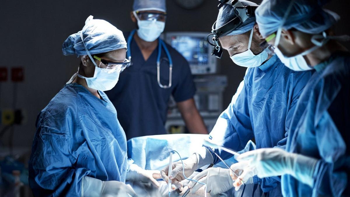
- Wide Local Excision: The primary treatment for phyllodes tumors is surgical removal. A wide local excision is performed to remove the tumor along with a margin of healthy tissue to reduce the risk of recurrence. The margin size depends on the tumor’s grade and characteristics.
- Lumpectomy: For smaller tumors with a low risk of recurrence, a lumpectomy (removal of the tumor with a small margin of surrounding tissue) may be sufficient.
- Mastectomy: In cases where the tumor is large, or if it has recurred multiple times, a mastectomy (removal of the entire breast) may be necessary. This approach is typically considered for tumors that are difficult to excise with clear margins.
2. Post-Surgical Treatment:
- Pathological Examination: After surgery, the removed tissue is examined to ensure that the tumor has been completely removed with clear margins. This helps determine if any additional treatment is necessary.
- Surveillance: Regular follow-up and surveillance are crucial to monitor for any signs of recurrence. This may include periodic physical examinations and imaging studies.
3. Radiation Therapy:
- Adjuvant Radiation: Radiation therapy is not routinely used for phyllodes tumors but may be considered in specific cases where the tumor has not been completely removed, or if it is high-grade and there is a high risk of recurrence. Radiation is generally reserved for situations where surgical margins are inadequate or if there is evidence of local recurrence.
4. Chemotherapy:
- Limited Use: Chemotherapy is not typically used for phyllodes tumors, as these tumors are usually resistant to systemic chemotherapy. It may be considered in cases of metastatic disease or if the tumor is aggressive and has spread beyond the primary site.
5. Hormone Therapy:
- Not Effective: Phyllodes tumors are generally not hormone-sensitive, so hormone therapy is not a standard treatment option.
Summary:
The mainstay of treatment for phyllodes tumors is surgical removal with wide local excision. The extent of surgery depends on the tumor’s size and characteristics. Post-surgical monitoring is essential to detect any recurrence early. In select cases, adjuvant radiation therapy may be used. Chemotherapy and hormone therapy are not typically effective for phyllodes tumors.

Leave a Reply
You must be logged in to post a comment.