What are the symptoms of photopsias?
Photopsias refer to the perception of flashes of light or visual disturbances in the absence of actual external light. Symptoms can vary based on the underlying cause but generally include:
1. Flashes of Light:
- Brief Flashes: Short bursts of light or “sparks” in the field of vision.
- Persistent Flashes: Continuous or recurring light sensations.
2. Visual Disturbances:
- Flashes of Color: Seeing bright colors or patterns, such as blue, red, or green flashes.
- Light Spots: Small, bright spots or “floaters” in the vision.
- Streaks or Lines: Perception of lines or streaks of light.
3. Other Associated Symptoms:
- Visual Field Defects: Areas of vision that are dim or absent, sometimes occurring alongside flashes.
- Blurred Vision: Difficulty focusing or seeing clearly during or after episodes.
- Eye Pain or Discomfort: In some cases, photopsias may be accompanied by eye pain or discomfort.
Possible Causes:
- Retinal Detachment: Flashes of light or visual disturbances may indicate a retinal tear or detachment.
- Migraine Headaches: Migraine-associated photopsias can occur as part of the aura phase of migraines.
- Vitreous Detachment: When the vitreous gel pulls away from the retina, it can cause flashes of light.
- Optic Neuritis: Inflammation of the optic nerve can lead to visual disturbances, including flashes of light.
- Ocular Migraines: Specific type of migraine affecting vision, often with light flashes.
- Other Eye Conditions: Conditions such as retinal damage or certain eye infections can also lead to photopsias.
Summary:
Photopsias are characterized by the perception of flashes of light or other visual disturbances without actual external light. These symptoms can be brief or persistent and may be associated with various eye or neurological conditions. If photopsias are experienced, especially if accompanied by other visual symptoms or sudden changes in vision, it is important to seek a comprehensive eye examination to determine the underlying cause and receive appropriate treatment.
What are the causes of photopsias?
Photopsias can be caused by a range of conditions affecting the eyes or the visual pathways in the brain. Here are some common causes:
1. Retinal Issues:
- Retinal Detachment: The separation of the retina from the underlying tissue can cause flashes of light.
- Retinal Tear: A tear in the retina can lead to sudden visual disturbances, including flashes.
- Vitreous Detachment: When the vitreous gel pulls away from the retina, it can cause flashes of light.
2. Migraine-Related Causes:
- Migraine Aura: Photopsias can occur as part of the visual aura associated with migraines. This can include flashes of light or zigzag patterns.
- Ocular Migraine: A type of migraine that affects vision, sometimes causing flashes of light or other visual disturbances.
3. Neurological Conditions:
- Optic Neuritis: Inflammation of the optic nerve can cause visual disturbances, including flashes of light.
- Brain Lesions: Tumors or lesions affecting the visual pathways in the brain can lead to photopsias.
4. Eye Conditions:
- Uveitis: Inflammation of the uvea (the middle layer of the eye) can sometimes lead to flashes or other visual disturbances.
- Glaucoma: Increased intraocular pressure and damage to the optic nerve can cause various visual symptoms, including flashes.
5. Systemic Conditions:
- Diabetic Retinopathy: Changes in the retina due to diabetes can cause visual disturbances, including flashes.
- Hypertensive Retinopathy: High blood pressure can affect the retina and lead to flashes or other visual symptoms.
6. Trauma:
- Eye Injury: Physical trauma to the eye can cause flashes of light or other visual disturbances.
7. Medication Side Effects:
- Certain Medications: Some medications may cause visual disturbances as a side effect, including flashes of light.
Summary:
Photopsias can result from various eye conditions, neurological disorders, or systemic diseases. Common causes include retinal detachment, migraines, optic neuritis, and trauma. Identifying the underlying cause requires a thorough eye examination and possibly additional neurological evaluations. If you experience photopsias, especially if they are sudden or accompanied by other symptoms, it is important to seek medical attention for an accurate diagnosis and appropriate treatment.
How is the diagnosis of photopsias made?
Diagnosing photopsias involves a combination of medical history, physical examination, and diagnostic tests to determine the underlying cause of the visual disturbances. Here’s a step-by-step approach to diagnosing photopsias:
1. Medical History:
- Symptom Description: Detailed information about the nature, duration, and frequency of the flashes of light or visual disturbances.
- Associated Symptoms: Inquiry about other symptoms such as headaches, blurred vision, or eye pain.
- Previous Medical History: Any history of eye conditions, neurological disorders, or systemic diseases.
- Medication Use: Review of current and past medications to identify possible side effects.
2. Physical Examination:
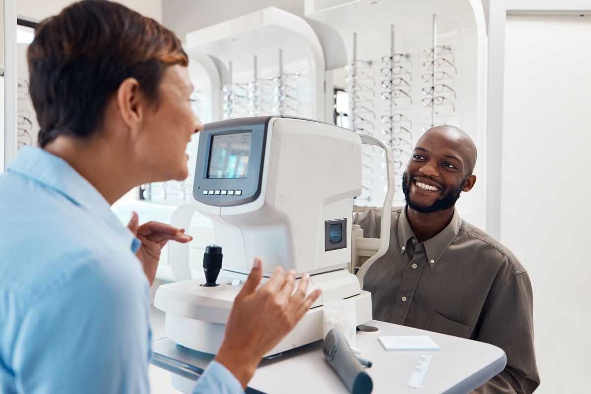
- Eye Examination: An eye specialist (ophthalmologist or optometrist) will conduct a thorough eye exam, including:
- Visual Acuity Test: Assessing how well you can see at various distances.
- Fundoscopy (Ophthalmoscopy): Examining the retina, optic nerve, and blood vessels to check for abnormalities.
- Slit-Lamp Examination: Using a microscope to examine the front structures of the eye and the retina.
3. Diagnostic Tests:
- Fundus Photography: Taking detailed images of the retina to identify any changes or abnormalities.
- Optical Coherence Tomography (OCT): Non-invasive imaging to view the layers of the retina and detect any retinal or macular issues.
- Fluorescein Angiography: Injecting a dye into the bloodstream and taking images of the retina to check for abnormalities in the blood vessels.
- Ultrasound: Using sound waves to examine the retina and vitreous body for signs of detachment or tears.
- Visual Field Test: Assessing the entire field of vision to detect any defects or changes.
4. Neurological Evaluation:
- Neurological Exam: If a neurological cause is suspected, a neurologist may perform a neurological examination to assess brain function and visual pathways.
- Magnetic Resonance Imaging (MRI): Imaging of the brain and optic pathways to detect any lesions, tumors, or other abnormalities.
- Computed Tomography (CT) Scan: May be used to evaluate the brain if there is a suspicion of trauma or other issues affecting vision.
5. Additional Tests:
- Blood Tests: To check for systemic conditions such as diabetes or hypertension that could affect the eyes.
- Electroretinography (ERG): Measures the electrical response of the retina to light stimulation, useful in diagnosing certain retinal conditions.
Summary:
Diagnosing photopsias involves a comprehensive approach including a detailed medical history, eye examination, and various diagnostic tests to identify the underlying cause. Identifying the specific cause is crucial for determining the appropriate treatment and management. If you experience photopsias, seeking prompt evaluation from an eye specialist or neurologist is important to ensure accurate diagnosis and effective treatment.
What is the treatment for photopsias?
The treatment for photopsias depends on the underlying cause. Here are general approaches to managing photopsias based on their cause:
1. Retinal Issues:
- Retinal Detachment or Tear: Treatment may involve laser therapy or cryotherapy to repair the retina. In some cases, surgery might be needed to reattach the retina.
- Vitreous Detachment: This condition often resolves on its own, but if it is causing significant symptoms or risk, monitoring and managing the condition is essential.
2. Migraine-Related Causes:
- Migraine Aura: Managing migraines with medications, such as triptans, anti-nausea drugs, or preventative treatments like beta-blockers, antidepressants, or antiepileptic drugs.
- Ocular Migraine: Avoiding known migraine triggers and using medications as prescribed by a healthcare provider.
3. Neurological Conditions:
- Optic Neuritis: Treatment may include corticosteroids to reduce inflammation. Addressing the underlying cause of optic neuritis, such as multiple sclerosis, is also important.
- Brain Lesions: Treatment depends on the specific type of lesion and may include surgery, radiation, or chemotherapy if the lesion is a tumor.
4. Eye Conditions:
- Uveitis: Treatment typically involves corticosteroids or other anti-inflammatory medications to reduce inflammation. Identifying and treating the underlying cause is also crucial.
- Glaucoma: Managing intraocular pressure with medications, laser therapy, or surgery to prevent damage to the optic nerve.
5. Systemic Conditions:
- Diabetic Retinopathy: Tight blood sugar control, laser treatment, or anti-VEGF injections to manage retinal changes.
- Hypertensive Retinopathy: Controlling blood pressure through lifestyle changes and medication to prevent further retinal damage.
6. Trauma:
- Eye Injury: Treatment may include medications for pain and inflammation, or surgery if there is significant damage.
7. Medication Side Effects:
- Adjusting Medications: If photopsias are caused by a medication, your healthcare provider may adjust the dosage or switch to an alternative medication.
General Management:
- Monitoring: Regular follow-up with an eye specialist to monitor the condition and adjust treatment as necessary.
- Lifestyle Modifications: Protecting your eyes from excessive light and reducing exposure to known triggers.
Summary:
Treatment for photopsias is tailored to the specific cause and may involve medical or surgical interventions, lifestyle adjustments, and regular monitoring. It’s important to work with healthcare providers to identify the underlying cause and develop an appropriate treatment plan. If you experience persistent or worsening photopsias, seeking evaluation from an eye specialist or other relevant healthcare provider is essential for proper management.

Leave a Reply
You must be logged in to post a comment.