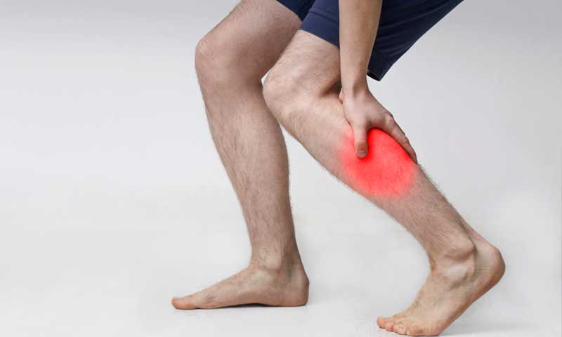
What are the symptoms of phlegmasia cerulea dolens?
Phlegmasia cerulea dolens is a severe and rare complication of deep vein thrombosis (DVT) characterized by significant venous obstruction and compromised blood flow. It is also known as “phlegmasia cerulea dolorosa” or “phlegmasia alba dolens” when it presents with different clinical features. The symptoms of phlegmasia cerulea dolens can be quite dramatic and severe, including:
1. Severe Pain:
- Intensity: Intense and debilitating pain in the affected limb.
- Location: Pain typically affects the leg where the thrombus (blood clot) is located.
2. Swelling:
- Extent: Profound swelling of the affected limb.
- Progression: Swelling may rapidly increase and involve the entire limb.
3. Cyanosis:
- Appearance: Bluish or purplish discoloration of the skin, often due to impaired venous return and reduced oxygenation of the blood.
- Severity: Cyanosis is a key feature distinguishing phlegmasia cerulea dolens from other types of DVT.
4. Temperature Changes:
- Coldness: The affected limb may feel cold to the touch compared to the unaffected limb.
- Temperature Changes: Differences in temperature can be observed, often cooler in the affected area.
5. Reduced Pulses:
- Pulses: Diminished or absent pulses in the affected limb, indicating impaired blood flow.
6. Skin Changes:
- Discoloration: The skin may exhibit a bluish or purplish hue due to reduced blood flow and oxygenation.
- Texture: The skin might also appear tense and shiny due to swelling.
7. Functional Impairment:
- Movement: Difficulty moving or using the affected limb due to pain and swelling.
8. Signs of Complications:
- Gangrene: In severe cases, prolonged and untreated phlegmasia cerulea dolens can lead to tissue necrosis or gangrene due to inadequate blood supply.
Summary:
Phlegmasia cerulea dolens presents with severe pain, profound swelling, cyanosis, coldness of the affected limb, reduced pulses, and potential skin changes. It is a medical emergency requiring prompt diagnosis and treatment to prevent serious complications such as tissue necrosis or gangrene.
What are the causes of phlegmasia cerulea dolens?
Phlegmasia cerulea dolens is a severe and rare complication of deep vein thrombosis (DVT) and is often caused by a combination of factors related to severe venous obstruction. The primary causes and contributing factors include:
1. Deep Vein Thrombosis (DVT):
- Main Cause: Phlegmasia cerulea dolens typically arises from a massive and extensive DVT in the deep veins of the leg. The thrombosis leads to significant obstruction of venous blood flow.
2. Venous Outflow Obstruction:
- Extent of Obstruction: The condition occurs when a large thrombus or multiple thrombi obstruct the major veins, such as the femoral, popliteal, or iliac veins. This obstruction leads to increased venous pressure and compromised blood flow.
3. Pre-existing Conditions:
- Cancer: Malignancies, particularly those associated with hypercoagulability or direct invasion of veins, can increase the risk of developing severe DVT and phlegmasia cerulea dolens.
- Pregnancy: Pregnancy and postpartum periods can increase the risk of thrombosis due to hormonal changes and increased venous pressure.
- Trauma or Surgery: Recent trauma or major surgical procedures, especially orthopedic surgeries or abdominal surgeries, can predispose individuals to severe DVT.
- Chronic Venous Insufficiency: Pre-existing chronic venous insufficiency can contribute to the development of severe complications related to DVT.
4. Hypercoagulable States:
- Blood Clotting Disorders: Conditions that increase the tendency for blood clot formation, such as inherited or acquired thrombophilia (e.g., factor V Leiden mutation, antiphospholipid syndrome), can lead to severe DVT and phlegmasia cerulea dolens.
5. Comorbidities:
- Heart Failure: Severe heart failure can contribute to venous congestion and increase the risk of DVT.
- Obesity: Obesity can exacerbate venous pressure and contribute to the development of severe DVT.
6. Catheter-Associated Thrombosis:
- Central Venous Catheters: Use of central venous catheters or other indwelling devices can increase the risk of thrombus formation and contribute to severe venous obstruction.
Summary:
Phlegmasia cerulea dolens is primarily caused by extensive DVT leading to severe venous obstruction. Contributing factors include cancer, pregnancy, recent trauma or surgery, chronic venous insufficiency, hypercoagulable states, comorbidities like heart failure, and catheter-associated thrombosis. Addressing the underlying cause and managing the severe obstruction is critical for treatment and prevention of complications.
How is the diagnosis of phlegmasia cerulea dolens made?
The diagnosis of phlegmasia cerulea dolens involves a combination of clinical evaluation, imaging studies, and sometimes laboratory tests. Here’s a step-by-step approach to diagnosing this condition:
1. Clinical Evaluation:
- Medical History:
- Symptoms Inquiry: The physician will ask about symptoms such as severe pain, swelling, and cyanosis in the affected limb.
- Risk Factors: Information about recent surgeries, trauma, pregnancy, cancer, or other risk factors for deep vein thrombosis (DVT).
- Physical Examination:
- Inspection: Assess for symptoms such as intense pain, significant swelling, cyanosis (bluish discoloration) of the limb, and coldness compared to the unaffected limb.
- Palpation: Check for tenderness, temperature differences, and diminished pulses in the affected limb.
- Assessment for Complications: Look for signs of complications like tissue necrosis.
2. Imaging Studies:
- Ultrasound:
- Use: The primary imaging tool for diagnosing DVT and evaluating the extent of venous thrombosis.
- Findings: May show large thrombi, deep vein thrombosis, and impaired venous flow.
- Venography:
- Use: An invasive imaging technique involving contrast dye to visualize the veins and confirm the presence and extent of thrombosis.
- Indication: Used if ultrasound findings are inconclusive or if more detailed imaging is needed.
- CT or MRI:
- Use: CT or MRI may be employed to assess the extent of thrombus and complications, especially if venography is not available or practical.
- Findings: Can show large thrombi, venous obstruction, and associated changes.
3. Laboratory Tests:
- D-dimer Test:
- Use: Measures the level of D-dimer, a substance released when a blood clot breaks down.
- Findings: Elevated D-dimer levels can indicate the presence of clotting, though it is not specific to phlegmasia cerulea dolens and can be elevated in other conditions.
- Blood Tests:
- Use: To evaluate underlying conditions such as hypercoagulable states or infections.
4. Differential Diagnosis:
- Rule Out Other Conditions:
- Complications of DVT: Ensure that symptoms are not due to other complications like cellulitis or arterial occlusion.
- Other Causes of Swelling: Differentiate from other causes of limb swelling and cyanosis.
Summary:
Diagnosing phlegmasia cerulea dolens involves a thorough clinical evaluation, imaging studies like ultrasound or venography, and laboratory tests to assess clotting and underlying conditions. The combination of severe pain, swelling, cyanosis, and imaging findings typically confirms the diagnosis. Prompt diagnosis is crucial for effective management and to prevent severe complications.
What is the treatment for phlegmasia cerulea dolens?
The treatment for phlegmasia cerulea dolens is focused on addressing the underlying deep vein thrombosis (DVT) and alleviating the symptoms and complications associated with the condition. The treatment approach typically includes the following steps:
1. Immediate Management:
- Anticoagulation Therapy:
- Purpose: To prevent further clot formation and allow the body’s natural thrombolytic processes to break down the existing clot.
- Medications: Initial treatment often involves intravenous heparin or low molecular weight heparin (LMWH), followed by oral anticoagulants such as warfarin or direct oral anticoagulants (DOACs) as per the patient’s needs.
- Thrombolysis:
- Purpose: To dissolve the thrombus more quickly and restore blood flow.
- Medications: Thrombolytic agents like tissue plasminogen activator (tPA) may be used in severe cases where there is a need for rapid clot resolution.
- Compression Therapy:
- Purpose: To reduce swelling and improve venous return.
- Method: Graduated compression stockings or devices may be used, although their application may be challenging in severe cases of phlegmasia cerulea dolens.
2. Surgical Interventions:
- Thrombectomy:
- Purpose: To physically remove the thrombus from the affected vein.
- Procedure: Surgical or catheter-directed thrombectomy may be performed in severe cases or if thrombolysis is not effective.
- Vena Cava Filter:
- Purpose: To prevent pulmonary embolism in patients who cannot receive anticoagulation therapy or in whom anticoagulation has failed.
- Indication: Placement of a filter in the inferior vena cava to catch any emboli before they reach the lungs.
3. Management of Complications:
- Pain Management:
- Medications: Analgesics and anti-inflammatory drugs may be prescribed to manage severe pain and discomfort.
- Wound Care:
- Monitoring: If tissue necrosis or ulcers develop, they need to be managed with appropriate wound care and possibly surgical intervention.
4. Long-Term Management:
- Anticoagulation Continuation:
- Duration: Long-term anticoagulation therapy may be required to prevent recurrence of DVT and manage the risk of complications.
- Monitoring and Follow-Up:
- Purpose: Regular follow-up with imaging studies to monitor the resolution of the thrombus and ensure effective management of the condition.
5. Addressing Underlying Causes:
- Treatment of Contributing Conditions:
- Cancer Management: If cancer is a contributing factor, appropriate cancer treatment should be coordinated.
- Pregnancy Management: If pregnancy is a factor, managing the condition in collaboration with obstetric care may be necessary.
Summary:
Treatment for phlegmasia cerulea dolens involves a combination of anticoagulation therapy, thrombolysis, and possibly surgical interventions to address the severe venous obstruction and restore blood flow. Managing pain, complications, and any underlying conditions is also crucial. Prompt and effective treatment is essential to prevent severe complications and improve patient outcomes.

Leave a Reply
You must be logged in to post a comment.