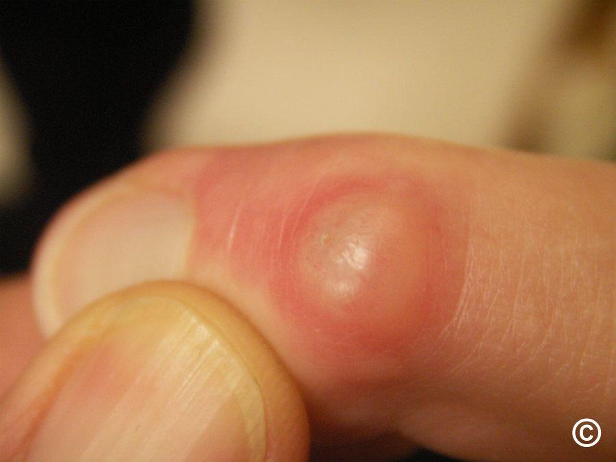
What are the symptoms of a myxoid cyst?
A myxoid cyst, also known as a digital mucous cyst or mucous pseudocyst, is a benign, fluid-filled lump that typically forms near the last joint of the fingers or toes, commonly affecting the dorsal surface of the digits. Myxoid cysts are associated with osteoarthritis and are often seen in middle-aged to older adults. Below are the typical symptoms:
1. Visible Lump:
- The most noticeable symptom is a small, smooth lump, usually located near the base of the fingernail or toenail on the distal interphalangeal joint (the last joint of the finger or toe).
- The cyst usually measures between 5 to 10 mm in diameter.
- It can be transparent, shiny, or slightly red in color.
2. Fluid-Filled Nature:
- The cyst contains a jelly-like, thick, clear fluid, which is the same fluid that lubricates the joints (synovial fluid).
- Occasionally, the cyst may burst, leading to the release of this clear, sticky fluid.
3. Nail Changes:
- A myxoid cyst may cause visible changes in the nearby nail. This can result in ridging, grooving, or splitting of the nail plate due to the pressure the cyst exerts on the nail matrix.
4. Discomfort or Pain:
- While many myxoid cysts are painless, some individuals may experience discomfort, tenderness, or pain, especially if the cyst is large or located in a place that is frequently irritated (e.g., due to pressure or trauma).
- Pain can also occur if the cyst presses against the joint or if it is associated with underlying osteoarthritis.
5. Decreased Range of Motion:
- In cases where the cyst is large or if it’s near a joint affected by osteoarthritis, it may limit the range of motion in the affected finger or toe, causing stiffness.
6. Secondary Infection:
- If the cyst bursts or is punctured, there is a risk of infection. Infected myxoid cysts can lead to redness, swelling, pain, and the discharge of pus.
- An infection can result in cellulitis or deeper infection in the surrounding tissues if not treated properly.
When to Seek Medical Attention:
- Persistent pain, nail deformities, or signs of infection (such as redness, heat, or discharge) should prompt medical evaluation.
- Some people may seek treatment for cosmetic reasons, especially if the cyst causes unsightly changes to the nail.
Myxoid cysts are typically harmless, but they can cause discomfort or cosmetic concerns depending on their size and location. Treatment options may include drainage, corticosteroid injections, or surgical removal, particularly if the cysts cause significant symptoms or cosmetic issues.
What are the causes of a myxoid cyst?
Myxoid cysts, also known as mucous cysts, are fluid-filled sacs that commonly occur in the fingers, particularly near the joints or at the base of the nails. While the exact cause of myxoid cysts is not fully understood, several factors are believed to contribute to their development:
1. Joint or Tendon Injury:
- Trauma or consistent pressure on the joints, particularly in the fingers, can lead to the formation of a myxoid cyst. This injury may result in the degeneration of the joint capsule or the tendon sheath, promoting cyst formation.
2. Arthritis:
- Conditions that lead to joint inflammation, such as osteoarthritis or rheumatoid arthritis, may increase the risk of developing myxoid cysts. Osteoarthritis, in particular, is frequently associated with these cysts, as it causes changes in joint structure and function.
3. Degenerative Changes:
- As people age, degenerative changes in the joints can occur, which might contribute to the development of myxoid cysts. These changes may result in the weakening of the joint capsule, allowing for the accumulation of mucous-like fluid.
4. Genetic Predisposition:
- There might be a genetic component that predisposes some individuals to develop myxoid cysts, although specific genetic factors are not well-defined.
5. Cystic Formation:
- Myxoid cysts are thought to arise from the accumulation of mucous fluid due to the degeneration of tissue around the joint or tendon sheath. This can create a pocket that fills with a gelatinous substance.
Conclusion:
While myxoid cysts are usually benign and may not require treatment unless symptomatic, understanding their potential causes can help in identifying risks and management strategies. If someone has concerns about a myxoid cyst, particularly if it is causing pain or affecting function, they should consult a healthcare professional for evaluation and discussion of possible treatment options.
How is the diagnosis of myxoid cyst made?
The diagnosis of a myxoid cyst is generally straightforward and involves a combination of clinical evaluation and, in some cases, imaging studies. Here are the steps typically involved in diagnosing a myxoid cyst:
1. Medical History:
- Symptom Assessment: The healthcare provider will take a detailed medical history, including information about the cyst’s onset, duration, and any associated symptoms such as pain or discomfort.
- Previous Injuries: The provider will inquire about any history of trauma or joint issues, as these factors can contribute to the development of myxoid cysts.
2. Physical Examination:
- Visual Inspection: During the physical examination, the clinician will visually inspect the lesion. Myxoid cysts typically present as smooth, round, or oval-shaped swellings on the dorsal aspect of the fingers or near the nails, often with a translucent or gelatinous appearance.
- Palpation: The doctor will palpate the cyst to assess its size, consistency, mobility, and tenderness. Myxoid cysts are usually firm but may have a soft, fluctuant quality due to their fluid content.
3. Differential Diagnosis:
- Exclusion of Other Conditions: The healthcare provider may consider other possible diagnoses that can present similarly, such as:
- Ganglion cysts
- Epidermoid cysts
- Pyogenic granulomas
- Other types of soft tissue tumors or lesions
- A thorough examination helps differentiate myxoid cysts from these other conditions.
4. Imaging Studies (if needed):
- Ultrasound: In some cases, an ultrasound may be performed to better visualize the cyst. It can help assess the lesion’s contents and characteristics, distinguishing it from other types of cysts or masses.
- X-ray: X-rays may be used if there are concerns about associated bone involvement or changes due to underlying arthritis.
5. Histological Examination (rarely needed):
- Biopsy: Typically, a biopsy is not required for diagnosis since myxoid cysts can often be diagnosed based on clinical presentation. However, in atypical cases or when malignancy is suspected, a fine needle aspiration (FNA) or excisional biopsy may be performed to obtain tissue for histological examination.
Conclusion:
Diagnosis of a myxoid cyst is usually made through a combination of patient history, physical examination, and potentially imaging studies. The characteristic appearance and location of the cyst typically allow for a confident diagnosis without the need for invasive procedures in most cases. If you’re experiencing symptoms of what you believe may be a myxoid cyst, it’s important to consult a healthcare provider for an accurate diagnosis and appropriate management options.
What is the treatment for a myxoid cyst?
The treatment for a myxoid cyst typically depends on several factors, including the size of the cyst, the symptoms it causes, and whether it recurs after any previous treatment. Common management options include:
1. Observation:
- Watchful Waiting: If the cyst is small, asymptomatic, and does not significantly affect function or appearance, a conservative approach of observation may be sufficient. Many myxoid cysts do not require intervention.
2. Aspiration:
- Needle Aspiration: In some cases, the cyst can be aspirated (drained) using a fine needle to remove the gelatinous fluid inside. This can provide temporary relief and improve appearance; however, myxoid cysts often recur after aspiration as the cyst cavity can refill with fluid.
3. Surgical Excision:
- Complete Removal: Surgical excision is the most definitive treatment for symptomatic myxoid cysts, particularly if they cause pain, discomfort, or functional impairment. During the procedure, the cyst and its capsule (lining) are removed to minimize the risk of recurrence.
- Cosmetic Reasons: Surgical intervention may also be performed for cosmetic reasons if the cyst is of significant concern to the patient.
4. Cryotherapy:
- Freezing the Cyst: In some instances, cryotherapy (freezing the cyst with liquid nitrogen) might be used to destroy the cyst tissue. This method may have varying success rates and is less commonly used.
5. Laser Therapy:
- Laser Options: Laser treatment may also be utilized for specific cases, although this approach is less common for myxoid cysts compared to other cosmetic or dermatological conditions.
6. Follow-Up Care:
- Monitoring Recurrence: Following treatment, especially after aspiration or excision, it’s important to monitor for recurrence. Some individuals may require additional treatment if the cyst returns.
Conclusion:
Most myxoid cysts are benign and either do not require treatment or can be managed conservatively. More invasive interventions, such as surgical excision, are recommended for symptomatic or recurrent cysts. If you suspect you have a myxoid cyst or are experiencing symptoms, it is advisable to consult a healthcare provider for an accurate diagnosis and tailored treatment recommendations.

Leave a Reply
You must be logged in to post a comment.