What are the symptoms of pneumomediastinum?
Pneumomediastinum, also known as mediastinal emphysema, occurs when air leaks into the mediastinum—the central compartment of the thoracic cavity that contains the heart, great vessels, trachea, esophagus, and other structures. The symptoms of pneumomediastinum can vary based on the severity of the condition and its underlying cause. Common symptoms include:
1. Chest Pain:
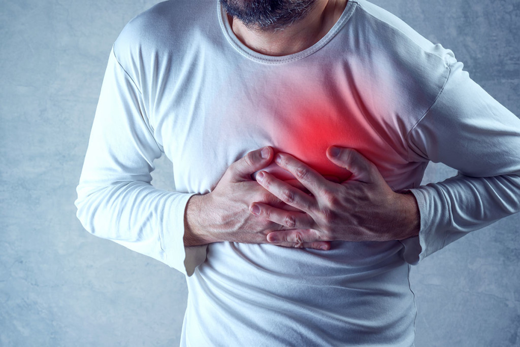
- Sharp or Dull Pain: The pain may be localized in the chest and can be sharp or dull, often described as a discomfort or pressure in the chest area.
2. Shortness of Breath:
- Difficulty Breathing: Patients may experience difficulty breathing or a feeling of breathlessness.
3. Cough:
- Persistent Cough: A cough may be present, sometimes accompanied by a feeling of irritation or soreness in the chest.
4. Neck Pain or Swelling:
- Subcutaneous Emphysema: Air trapped in the mediastinum can sometimes spread to the neck, causing swelling or a sensation of fullness. This can be noticeable as swelling or crepitus (a crackling sensation) under the skin.
5. Hoarseness:
- Voice Changes: Involvement of the recurrent laryngeal nerve can cause hoarseness or changes in the voice.
6. Difficulty Swallowing:
- Dysphagia: Swelling or pressure in the mediastinum may cause difficulty swallowing or a sensation of pressure in the throat.
7. Respiratory Distress:
- Severe Cases: In severe cases, pneumomediastinum can lead to respiratory distress and more pronounced difficulty breathing.
8. Systemic Symptoms:
- Fever or Malaise: In cases where the pneumomediastinum is associated with infection or underlying illness, systemic symptoms such as fever or general malaise may be present.
The symptoms of pneumomediastinum can overlap with other conditions affecting the chest, so a thorough evaluation by a healthcare professional is necessary to determine the cause and appropriate treatment.
What are the causes of pneumomediastinum?
Pneumomediastinum occurs when air enters the mediastinum, the central compartment of the thoracic cavity. Several conditions and activities can lead to the development of pneumomediastinum:
1. Barotrauma:
- Rapid Changes in Air Pressure: Activities or conditions that involve rapid changes in air pressure, such as scuba diving, flying, or mechanical ventilation, can cause barotrauma (damage from pressure changes) and lead to pneumomediastinum.
2. Chest Trauma:
- Injury: Direct trauma to the chest, such as from a car accident or a fall, can cause rupture of the airways or lung tissue, leading to the accumulation of air in the mediastinum.
3. Esophageal Rupture:
- Trauma or Vomiting: A rupture or tear in the esophagus, often due to severe vomiting or trauma, can allow air to escape into the mediastinum.
4. Respiratory Conditions:
- Severe Coughing: Intense coughing, especially from conditions like whooping cough or chronic bronchitis, can cause pneumomediastinum by increasing intra-thoracic pressure.
- Asthma: Severe asthma attacks with high pressure and forced breathing can also contribute.
5. Spontaneous Pneumomediastinum:
- Idiopathic: Sometimes, pneumomediastinum occurs spontaneously without an obvious underlying cause, particularly in young adults and adolescents.
6. Medical Procedures:
- Endoscopy: Certain medical procedures, such as endoscopy or bronchoscopy, can introduce air into the mediastinum inadvertently.
- Mechanical Ventilation: High pressure settings on mechanical ventilators can sometimes cause air to escape into the mediastinum.
7. Connective Tissue Disorders:
- Marfan Syndrome: Some connective tissue disorders, like Marfan syndrome, may increase the risk of pneumomediastinum due to weakened tissues and increased susceptibility to rupture.
8. Infections:
- Infection-Induced Inflammation: Certain infections, such as those causing mediastinitis (inflammation of the mediastinum), may lead to the accumulation of air in this area.
9. Overexertion:
- Heavy Lifting or Straining: Activities that involve heavy lifting or intense physical exertion can increase the risk of pneumomediastinum due to the resulting pressure changes in the thoracic cavity.
Identifying the underlying cause of pneumomediastinum is crucial for effective management and treatment. If you suspect pneumomediastinum or experience symptoms such as chest pain or difficulty breathing, it’s important to seek medical evaluation promptly.
How is the diagnosis of pneumomediastinum made?
The diagnosis of pneumomediastinum involves a combination of clinical evaluation, imaging studies, and sometimes additional diagnostic procedures. Here’s how it is typically diagnosed:
1. Clinical Evaluation:
- Medical History: The healthcare provider will review the patient’s medical history, including any recent activities, injuries, or symptoms that might suggest pneumomediastinum.
- Physical Examination: The examination may reveal signs such as neck swelling, subcutaneous emphysema (air under the skin), or tenderness in the chest area.
2. Imaging Studies:
- Chest X-ray: This is often the first imaging test performed. A chest X-ray can reveal the presence of air in the mediastinum as well as other associated signs, such as air around the lungs (pneumothorax) or fluid accumulation.
- CT Scan (Computed Tomography): A CT scan of the chest is more sensitive and specific for diagnosing pneumomediastinum. It provides detailed images of the mediastinum and can confirm the presence of air, assess the extent of the condition, and help identify potential causes, such as esophageal rupture or trauma.
3. Additional Diagnostic Procedures:
- Esophagram (Barium Swallow Study): If esophageal rupture is suspected, a barium swallow study may be performed. This involves ingesting a contrast material (barium) to visualize the esophagus and identify any tears or leaks.
- Bronchoscopy: In cases where the cause is unclear and involves the airways, a bronchoscopy may be performed to directly visualize the trachea and bronchi.
4. Laboratory Tests:
- Blood Tests: While not diagnostic for pneumomediastinum itself, blood tests can help assess overall health and identify any underlying infections or inflammation that might be contributing to the condition.
5. Clinical Observation:
- Monitoring Symptoms: In some cases, if the diagnosis is uncertain, monitoring symptoms over time and observing for changes may provide additional clues.
The combination of these methods helps to confirm the presence of pneumomediastinum, determine its cause, and guide appropriate treatment. If you experience symptoms such as chest pain, difficulty breathing, or swelling in the neck, it’s important to seek medical attention promptly for evaluation and diagnosis.
What is the treatment for pneumomediastinum?
The treatment for pneumomediastinum depends on the underlying cause, severity of symptoms, and overall health of the patient. In many cases, the condition resolves on its own, but management may be necessary to address symptoms and prevent complications. Here’s a general approach to treatment:
1. Observation and Supportive Care:
- Rest and Monitoring: For mild cases of pneumomediastinum, especially when symptoms are minimal and there is no significant underlying cause, observation and rest are often sufficient. Patients are monitored for any worsening of symptoms.
- Pain Management: Over-the-counter pain relievers, such as acetaminophen or ibuprofen, can help manage chest pain or discomfort.
2. Addressing Underlying Causes:
- Treating the Cause: If an underlying condition or activity caused the pneumomediastinum (e.g., severe coughing, esophageal rupture, or trauma), treatment will focus on managing that specific issue. For instance:
- Esophageal Rupture: Requires prompt surgical intervention and supportive care, including antibiotics to prevent infection.
- Trauma: May need surgical repair or other interventions depending on the extent of the injury.
- Respiratory Conditions: Treatment may involve managing the respiratory condition that contributed to the pneumomediastinum.
3. Medications:
- Antibiotics: If there is an associated infection or risk of infection, antibiotics may be prescribed.
- Corticosteroids: In some cases, corticosteroids may be used to reduce inflammation, especially if there is significant swelling or inflammation in the mediastinum.
4. Medical Procedures:
- Drainage: In rare cases where there is a large amount of air or associated complications, procedures to remove the air (e.g., needle aspiration) may be necessary. This is usually considered only if there are severe symptoms or complications.
5. Avoiding Aggravating Factors:
- Avoid Activities: Patients are often advised to avoid activities that could increase intrathoracic pressure, such as heavy lifting or vigorous physical activity, until the condition resolves.
6. Follow-Up Care:
- Regular Monitoring: Follow-up visits may be necessary to monitor the resolution of pneumomediastinum and ensure that symptoms are improving.
7. Emergency Care:
- Severe Cases: If pneumomediastinum is associated with severe symptoms or complications, such as difficulty breathing or significant chest pain, emergency medical care is required.
Treatment is tailored to the individual based on the severity of the condition, the underlying cause, and the overall health of the patient. If you suspect pneumomediastinum or experience symptoms like chest pain or difficulty breathing, seek medical evaluation to determine the appropriate course of action.

Leave a Reply
You must be logged in to post a comment.