What is involved in a PET scan?
A PET (Positron Emission Tomography) scan is a diagnostic imaging technique that provides detailed images of the body’s metabolic processes. It is often used to detect and monitor diseases, including cancers, heart conditions, and neurological disorders. Here’s an overview of what’s involved in a PET scan:
1. Preparation:
- Dietary Instructions:
- Fasting: Patients may be required to fast for several hours before the scan, typically 4 to 6 hours, to ensure accurate results. This is because food and drink can affect the distribution of the radiotracer used in the scan.
- Medication:
- Consultation: Inform the healthcare provider of any medications you are taking, as some might need to be paused or adjusted before the scan.
- Clothing:
- Comfortable Clothing: Wear loose, comfortable clothing. You may be asked to change into a hospital gown for the scan.
2. Radiotracer Injection:
- Radiotracer:
- Type: A small amount of a radioactive substance (radiotracer) is injected into a vein, usually in your arm. The radiotracer is typically a form of glucose or another compound that accumulates in areas of high metabolic activity.
- Uptake Time:
- Waiting Period: After the injection, you will wait for a period (usually about 30 to 60 minutes) to allow the radiotracer to be absorbed by the body tissues and organs. During this time, it’s important to remain still and avoid strenuous activities.
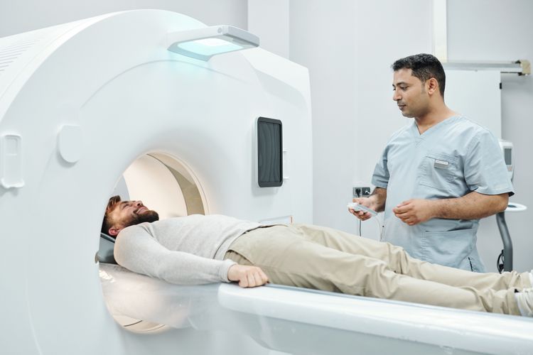
3. Scanning Procedure:
- Positioning:
- Scan Position: You will lie on a table that slides into the PET scanner. The scanner is a large, cylindrical machine that detects the radiation emitted by the radiotracer in your body.
- Scanning:
- Duration: The scanning process usually takes about 20 to 30 minutes, depending on the area being studied. The scanner will move slowly around your body, capturing images from multiple angles.
- Instructions: You may be asked to remain still and hold your breath briefly while the scan is being performed to ensure clear images.
4. Post-Scan:
- Radiotracer Elimination:
- Excretion: The radioactive substance will gradually leave your body through urine, feces, and sweat. Drinking plenty of fluids can help speed up the elimination process.
- Activity:
- Normal Activities: You can usually resume normal activities immediately after the scan, although you may be advised to drink extra fluids to help flush out the radiotracer.
5. Results and Follow-Up:
- Image Analysis:
- Interpretation: A radiologist or nuclear medicine specialist will analyze the images and provide a report based on the findings.
- Consultation: Your healthcare provider will discuss the results with you and determine any necessary follow-up steps or treatments based on the findings of the PET scan.
Summary:
A PET scan involves the injection of a radiotracer, waiting for the tracer to accumulate in the body, and then imaging with a PET scanner. The procedure is non-invasive and typically well-tolerated, providing valuable information about the metabolic activity and function of tissues and organs. It is commonly used in the diagnosis and management of various conditions, including cancer, heart disease, and neurological disorders.
What diseases and conditions can a PET scan detect?
A PET (Positron Emission Tomography) scan is a powerful imaging tool that helps detect and monitor a range of diseases and conditions by evaluating the metabolic activity of tissues and organs. Here are some of the key diseases and conditions that a PET scan can help detect:
1. Cancer:
- Detection and Diagnosis:
- Primary Cancers: Identifies and locates primary tumors (e.g., lung cancer, breast cancer, lymphoma).
- Metastasis: Detects cancer spread to other parts of the body.
- Recurrence: Monitors for cancer recurrence after treatment.
- Types of Cancer Detected:
- Lung Cancer
- Breast Cancer
- Colorectal Cancer
- Lymphomas
- Melanoma
- Prostate Cancer
2. Cardiovascular Diseases:
- Heart Conditions:
- Coronary Artery Disease (CAD): Evaluates the extent of damage to the heart muscle and assesses the effectiveness of treatment.
- Myocardial Viability: Assesses the viability of heart tissue, especially after a heart attack.
3. Neurological Disorders:
- Brain Conditions:
- Alzheimer’s Disease: Identifies changes in brain metabolism associated with early Alzheimer’s disease.
- Parkinson’s Disease: Evaluates brain function and helps differentiate Parkinson’s disease from other movement disorders.
- Seizure Disorders: Helps localize the area of the brain responsible for seizure activity.
4. Infectious Diseases:
- Infection Localization:
- Abscesses: Identifies the presence and location of infections or abscesses.
- Infectious Diseases: Evaluates the spread of certain infections, particularly those resistant to standard imaging techniques.
5. Inflammatory Diseases:
- Chronic Inflammation:
- Sarcoidosis: Detects and monitors the progression of sarcoidosis, an inflammatory disease affecting multiple organs.
- Vasculitis: Evaluates inflammation of blood vessels associated with various forms of vasculitis.
6. Endocrine Disorders:
- Thyroid Conditions:
- Thyroid Cancer: Assesses thyroid function and detects cancerous growths.
- Hyperthyroidism/Hypothyroidism: Evaluates thyroid activity and functionality.
7. Trauma and Injury:
- Assessment:
- Traumatic Brain Injury: Evaluates brain function and metabolism following traumatic injuries.
Summary:
A PET scan is useful for diagnosing, staging, and monitoring a wide range of diseases, including cancers, cardiovascular conditions, neurological disorders, infectious diseases, and inflammatory conditions. It provides valuable information about metabolic and functional changes in tissues, which can complement other imaging modalities and clinical evaluations. If you have specific concerns or conditions, your healthcare provider can determine if a PET scan is appropriate for your diagnosis and treatment plan.
There are many kinds of scans – MRI, CT, PET, Etc – which scan is best at detecting cancers before they become symptomatic?
Each type of imaging scan (MRI, CT, PET, etc.) serves different purposes in detecting and diagnosing cancer. The best scan for detecting cancer before it becomes symptomatic depends on the type of cancer, its location, and what is being evaluated. Here’s an overview of common imaging techniques used for early cancer detection:
1. MRI (Magnetic Resonance Imaging)
- Strength: Provides highly detailed images of soft tissues and organs without radiation.
- Best for: Detecting brain, spinal cord, breast, liver, prostate, and musculoskeletal cancers.
- Screening Use: MRI is often used in high-risk patients, such as those with a family history of breast cancer. It is particularly sensitive for early detection of breast cancer in dense breast tissue.
2. CT (Computed Tomography) Scan
- Strength: Uses X-rays to create detailed cross-sectional images of the body.
- Best for: Detecting cancers in organs such as the lungs, liver, pancreas, and colon.
- Screening Use: Low-dose CT (LDCT) scans are used for early detection of lung cancer in high-risk individuals (e.g., long-term smokers).
3. PET (Positron Emission Tomography) Scan
- Strength: Shows metabolic activity of tissues, which can indicate rapidly growing cancer cells.
- Best for: Detecting cancers that are metabolically active, such as lymphomas, and identifying cancer spread (metastasis).
- Screening Use: Not typically used for screening, but combined PET/CT scans can detect certain cancers and evaluate their activity at an early stage.
4. Mammography
- Strength: Uses low-dose X-rays to detect abnormalities in breast tissue.
- Best for: Early detection of breast cancer, especially in women over the age of 40.
- Screening Use: A standard tool for routine breast cancer screening in women, often detecting tumors before they cause symptoms.
5. Ultrasound
- Strength: Uses sound waves to create images, effective for soft tissues and organs.
- Best for: Detecting cancers of the liver, kidneys, and reproductive organs.
- Screening Use: Often used to evaluate suspicious lumps, particularly in the thyroid, testicles, or breasts.
6. Bone Scans
- Strength: Uses radioactive tracers to detect areas of increased bone activity.
- Best for: Detecting bone metastases or primary bone cancers.
- Screening Use: Used more often to detect cancer spread than for initial cancer detection.
Which Scan is Best for Early Detection?
- MRI and mammography are commonly used for early detection in high-risk individuals for certain cancers (e.g., breast and prostate cancer).
- Low-dose CT scans are effective for early detection of lung cancer in high-risk populations.
- PET scans are useful for detecting cancer spread but not typically used as a primary screening tool for early detection.
Each cancer type and risk factor may call for a different imaging technique, so consultation with a healthcare provider is crucial to determine the most appropriate scan for early detection based on individual risk factors.
Leave a Reply
You must be logged in to post a comment.