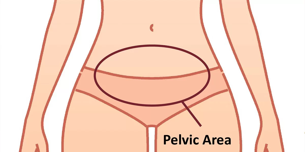
What are the symptoms of pelvic fractures?
Pelvic fractures can cause a variety of symptoms depending on the severity of the injury. Common symptoms of pelvic fractures include:
1. Pain
- Severe pain in the hip or pelvis: Pain is typically sharp and worsens with movement or weight-bearing activities.
- Lower back or abdominal pain: Pain may radiate to the lower back or abdomen.
2. Difficulty Walking or Standing
- Inability to walk: Severe fractures may make walking or even standing difficult or impossible.
- Pain while standing or sitting: Even simple movements like sitting or standing can cause significant discomfort.
3. Bruising and Swelling
- Bruising (ecchymosis): Visible bruising around the pelvis, groin, or thighs.
- Swelling: The pelvic region may appear swollen due to internal bleeding or inflammation.
4. Leg Symptoms
- Leg weakness or numbness: Nerve involvement can cause tingling, numbness, or weakness in the legs.
- One leg appearing shorter: In severe cases, one leg may appear shorter due to displacement of the pelvic bones.
5. Difficulty Urinating or Bowel Movements
- Urinary issues: Difficulty urinating, blood in the urine (hematuria), or an inability to control bladder function may indicate damage to the bladder or urethra.
- Bowel issues: Difficulty with bowel movements or control can occur if there is damage to the rectum or surrounding structures.
6. Internal Bleeding
- Signs of shock: In severe fractures, internal bleeding may occur, leading to symptoms of shock such as dizziness, fainting, rapid pulse, low blood pressure, and pale, cool, clammy skin.
- Abdominal distension: The abdomen may appear swollen due to internal bleeding.
7. Hip or Groin Deformity
- Visible deformity: The hip or pelvis may appear misshapen or misaligned in more severe fractures.
Conclusion
Pelvic fractures can be serious and require immediate medical attention, especially if symptoms such as difficulty walking, urinary issues, or signs of shock are present. Early diagnosis and treatment are critical for proper healing and to prevent complications. If a pelvic fracture is suspected, it is important to seek emergency medical care.
What are the causes of pelvic fractures?
Pelvic fractures are typically caused by significant trauma or stress to the pelvic area. Here are the primary causes:
1. High-Energy Trauma
- Motor Vehicle Accidents: The most common cause of severe pelvic fractures, often involving high-speed collisions.
- Falls from Heights: Falling from a significant height can cause a major impact on the pelvis, leading to fractures.
- Crush Injuries: Situations where the pelvis is subjected to heavy pressure, such as industrial or construction accidents.
2. Low-Energy Trauma (Common in Older Adults)
- Falls in the Elderly: In older adults, especially those with osteoporosis, even a minor fall from standing height can lead to a pelvic fracture.
- Weakening of Bones due to Osteoporosis: Bone thinning in older adults increases the risk of fractures with minimal trauma.
3. Stress Fractures
- Repetitive Strain or Overuse: Athletes, particularly runners or military personnel, can develop stress fractures in the pelvis due to repetitive stress on the bones.
- Insufficient Bone Healing: Recurrent strain without adequate rest can lead to microfractures that eventually result in a full fracture.
4. Bone Diseases
- Bone Cancer or Tumors: Malignant or benign tumors can weaken the pelvic bones, making them more susceptible to fractures.
- Paget’s Disease of Bone: This chronic disorder causes bones to become deformed and weakened, increasing the risk of fractures.
5. Congenital or Genetic Conditions
- Osteogenesis Imperfecta: This genetic disorder, also known as brittle bone disease, results in bones that are more prone to fractures, including the pelvis.
Conclusion
Pelvic fractures are usually the result of significant trauma, especially in younger individuals, or weakened bones due to conditions like osteoporosis in older adults. Prevention and treatment depend on addressing underlying risk factors such as fall prevention, bone health, and trauma management.
How is the diagnosis of pelvic fractures made?
Diagnosing pelvic fractures involves a combination of clinical evaluation, imaging studies, and sometimes laboratory tests. The process typically includes:
1. Medical History and Physical Examination:
- Patient History: The healthcare provider will take a detailed history of the injury, including the mechanism of trauma (e.g., fall, motor vehicle accident), onset of symptoms, and any pre-existing conditions that may affect bone health.
- Physical Examination: A thorough physical exam is conducted to assess for signs of pelvic fractures, including pain, tenderness, swelling, bruising, deformity, and the ability to move. The exam may also include checking for stability of the pelvis and evaluating neurovascular status (e.g., sensation, blood flow).
2. Imaging Studies:
- X-rays: The first-line imaging study to evaluate pelvic fractures. X-rays can reveal the presence, location, and extent of the fracture.
- CT (Computed Tomography) Scan: Provides detailed cross-sectional images of the pelvis, which are helpful for assessing complex fractures, detecting subtle fractures not seen on X-rays, and evaluating the involvement of internal organs or blood vessels.
- MRI (Magnetic Resonance Imaging): Used in certain cases to assess soft tissue injuries, bone marrow edema, or occult fractures that are not visible on X-rays or CT scans. MRI is also helpful in identifying injuries to ligaments, muscles, and other soft tissues.
- Ultrasound: Occasionally used to assess soft tissue injuries or hematomas in the pelvic area, particularly in emergency settings.
3. Laboratory Tests:
- Blood Tests: May be ordered to assess for internal bleeding, anemia, or infection. Tests might include complete blood count (CBC), blood type and crossmatch, and coagulation profile.
- Urinalysis: To check for blood in the urine, which can indicate trauma to the urinary tract.
4. Clinical Assessments:
- Neurovascular Examination: To assess nerve function and blood flow in the lower extremities, checking for symptoms like numbness, tingling, or decreased pulse.
- Pelvic Stability Tests: Physical maneuvers performed by the healthcare provider to assess the stability of the pelvic ring, though these are done cautiously to avoid further injury.
5. Specialized Diagnostic Procedures:
- Angiography: In cases of suspected vascular injury, angiography may be used to visualize blood vessels and identify sources of bleeding.
- Cystourethrogram: A contrast study used to evaluate the bladder and urethra if there is concern for urinary tract injuries.
6. Consultations and Multidisciplinary Team Involvement:
- In complex cases, a multidisciplinary team including orthopedic surgeons, trauma surgeons, radiologists, and other specialists may be involved in the diagnosis and management.
Summary:
The diagnosis of pelvic fractures is typically made based on clinical findings and confirmed with imaging studies. The choice of imaging modality depends on the suspected severity and complexity of the fracture. Accurate diagnosis is crucial for determining the appropriate treatment plan and managing potential complications.
What is the treatment for pelvic fractures?
The treatment for pelvic fractures depends on the severity and type of the fracture, the specific bones involved, the stability of the pelvic ring, and the overall condition of the patient. The primary goals of treatment are to relieve pain, ensure proper healing of the bones, restore function, and prevent complications. Treatment options can range from conservative management to surgical intervention. Here are the main approaches:
1. Conservative Management:
- Rest and Activity Modification: Patients may be advised to rest and avoid weight-bearing activities to allow the fracture to heal. Bed rest or limited mobility may be necessary in the initial phase.
- Pain Management: Pain relief is a crucial component, often involving medications such as nonsteroidal anti-inflammatory drugs (NSAIDs), acetaminophen, or opioids for severe pain.
- Physical Therapy: Once the acute pain subsides, physical therapy may be introduced to help restore mobility, strengthen muscles, and improve overall function.
- Bracing and Supports: In some cases, external supports such as pelvic binders or braces may be used to stabilize the pelvis.
2. Minimally Invasive Procedures:
- Angiographic Embolization: If there is significant bleeding, angiography followed by embolization can be performed to control hemorrhage by blocking the bleeding vessels.
- Percutaneous Fixation: In some cases, minimally invasive techniques, such as the insertion of screws or rods through small incisions, can stabilize the fracture without open surgery.
3. Surgical Treatment:
- Open Reduction and Internal Fixation (ORIF): This involves surgical exposure of the fracture site to realign the bones and stabilize them with plates, screws, or rods. ORIF is commonly used for unstable fractures or those involving multiple breaks.
- External Fixation: A frame outside the body is connected to the pelvis with pins, providing stability while the bones heal. This method is often used in emergencies or when internal fixation is not feasible.
- Debridement and Repair: In cases where there is associated soft tissue injury or open fractures, surgical debridement (cleaning out dead tissue) and repair may be necessary.
4. Rehabilitation and Recovery:
- Physical Therapy and Rehabilitation: As healing progresses, a structured rehabilitation program is essential to regain strength, flexibility, and mobility. Physical therapists work with patients to develop personalized exercise routines.
- Occupational Therapy: Helps patients adapt to daily activities and improve their quality of life during recovery.
5. Management of Complications:
- Treatment of Associated Injuries: If other injuries (e.g., to the bladder, urethra, nerves) are present, they will be addressed as part of the treatment plan.
- Prevention of Deep Vein Thrombosis (DVT): Immobilization can increase the risk of blood clots, so blood thinners or compression devices may be used to prevent DVT.
- Infection Control: In cases of open fractures or surgical interventions, antibiotics may be administered to prevent infection.
6. Long-Term Management:
- Follow-Up Care: Regular follow-up appointments with healthcare providers to monitor healing and adjust treatment plans as needed.
- Chronic Pain Management: For some patients, chronic pain may persist and require ongoing management.
Summary:
The treatment of pelvic fractures is individualized based on the specific characteristics of the fracture and the patient’s overall health. While many fractures can be managed conservatively, severe or unstable fractures often require surgical intervention. Comprehensive care, including pain management, physical therapy, and monitoring for complications, is essential for optimal recovery.

Leave a Reply
You must be logged in to post a comment.