What are the symptoms of osteoradionecrosis?
The symptoms of osteoradionecrosis (ORN), a condition where bone tissue dies due to radiation therapy, can vary depending on the severity and location, often affecting the jaw (mandible) after radiation treatment for head and neck cancers. Key symptoms include:
- Pain: Persistent or worsening pain in the affected bone area.
- Swelling: Swelling of the tissues around the bone.
- Exposed bone: Visible bone through an opening in the skin or gums, often after trauma or dental procedures.
- Infection: Recurrent or chronic infections around the affected area.
- Jaw stiffness: Difficulty opening the mouth or jaw (trismus).
- Loose teeth: Teeth in the area of the dead bone may become loose or fall out.
- Drainage: Pus or other discharge from the site of exposed bone.
- Delayed healing: Wounds or sores in the area may heal very slowly or not at all.
- Fractures: In advanced cases, spontaneous fractures of the bone may occur due to its weakened state.
Early diagnosis and treatment are essential to manage osteoradionecrosis and prevent more severe complications.
What are the causes of osteoradionecrosis?
Osteoradionecrosis (ORN) occurs when bone tissue dies as a result of radiation therapy, most often following treatment for cancers in the head and neck region. The main causes of ORN include:
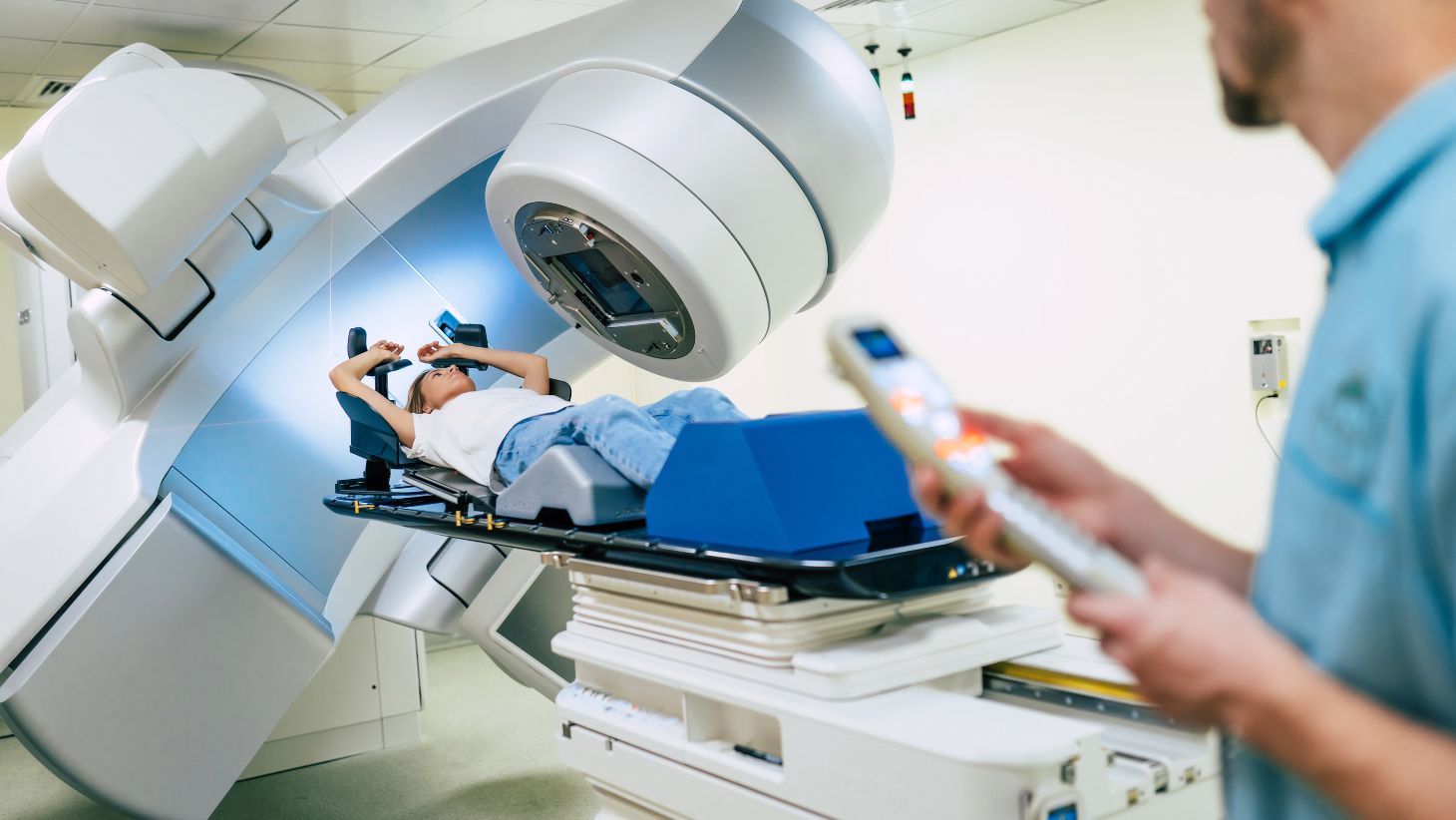
- Radiation therapy: High doses of radiation used to treat cancer can damage the blood vessels that supply the bone, leading to reduced blood flow, poor healing, and eventually bone death. ORN commonly affects the jawbone (mandible) due to its proximity to radiation fields in head and neck cancer treatments.
- Trauma or dental procedures: Procedures like tooth extractions or surgeries after radiation therapy can trigger ORN in the irradiated bone. The bone’s reduced healing capacity makes it more vulnerable to trauma.
- Infection: Radiation can compromise the bone’s ability to fight infection, increasing the risk of osteoradionecrosis if an infection occurs after dental or surgical procedures.
- High radiation doses: Higher doses of radiation, especially when used in conjunction with chemotherapy, increase the risk of ORN.
- Poor oral health: Pre-existing dental or gum disease before radiation treatment can increase the likelihood of developing ORN. Poor oral hygiene or untreated dental issues in irradiated areas may lead to infections that cause bone damage.
- Smoking and alcohol use: These factors reduce blood flow to the tissues and may further impair healing, raising the risk of developing ORN.
ORN is a complication that develops over time, often months or years after radiation therapy, and can be influenced by these contributing factors.
How is the diagnosis of osteoradionecrosis made?
The diagnosis of osteoradionecrosis (ORN) involves a combination of clinical evaluation, imaging studies, and, in some cases, biopsy to assess the extent of bone damage. Here’s how the diagnosis is typically made:
- Clinical examination: The doctor or dentist will examine the affected area, looking for key signs of ORN such as exposed bone, non-healing wounds, infections, or pus discharge. The patient’s medical history, particularly previous radiation therapy to the area, is crucial for diagnosis.
- Imaging studies:
- X-rays: Initial imaging to evaluate bone damage and the extent of necrosis.
- CT scan: Provides more detailed information about bone and surrounding tissue, including the degree of bone destruction.
- MRI: Helpful in assessing soft tissue involvement and infection but less commonly used than CT scans for bone assessment.
- Bone scans: Can help detect areas of reduced bone activity, suggesting necrosis or bone death.
- Biopsy (if needed): In some cases, a biopsy of the affected bone may be taken to rule out other conditions, such as cancer recurrence or bone infection (osteomyelitis), which may mimic ORN.
- Microbiological testing: If infection is present, pus or discharge from the affected area may be tested to identify any bacteria, guiding appropriate treatment.
The combination of a patient’s radiation history, clinical symptoms, and imaging findings is usually sufficient for diagnosing osteoradionecrosis.
What is the treatment for osteoradionecrosis?
The treatment of osteoradionecrosis (ORN) aims to alleviate symptoms, control infection, promote healing, and, when possible, preserve the integrity of the affected bone and surrounding tissues. The management of ORN is multidisciplinary and may involve oral and maxillofacial surgeons, medical doctors, dental professionals, and other healthcare providers. Here’s an overview of the treatment options:
1. Conservative Management:
- Antibiotics: If there is an infection present, antibiotics may be prescribed to control the infection. The choice of antibiotic will depend on the suspected or identified pathogens.
- Analgesics: Pain management is crucial, and non-steroidal anti-inflammatory drugs (NSAIDs) or opioids may be used to alleviate pain symptoms.
- Oral Hygiene: Improved oral hygiene and regular dental care are essential in managing ORN. Antiseptic mouthwashes may be recommended to prevent infections.
- Mucosal Care: Lesions may be treated with topical agents or dressings to promote healing and alleviate discomfort.
2. Surgical Intervention:
- Debridement: Surgical debridement of necrotic bone and infected tissue may be necessary. This procedure aims to remove devitalized tissue to encourage healing and prevent the spread of infection.
- Bone Grafting: In cases where considerable bone loss has occurred, bone grafting may be performed to reconstruct the affected area. This can involve using bone from the patient (autograft) or synthetic materials.
- Reconstructive Surgery: For more advanced cases, reconstructive surgery may involve the use of vascularized bone flaps (transplanting living bone with its blood supply) to restore the affected area’s structure and function.
3. Hyperbaric Oxygen Therapy (HBOT):
- HBOT has been used as an adjunctive treatment for ORN. It involves delivering 100% oxygen at increased atmospheric pressure, which can enhance oxygen supply to damaged tissues, improve healing, reduce tissue edema, and potentially promote angiogenesis (new blood vessel formation).
4. Management of Complications:
- Managing complications such as osteomyelitis (infection of the bone) or severe infections may require targeted surgical interventions and additional medical therapies.
5. Patient Education and Support:
- Providing education on maintaining oral hygiene, managing dental health, and recognizing early signs of ORN or complications is an essential part of treatment. Support from healthcare providers, nutritionists, and mental health professionals may also be beneficial.
Conclusion:
The treatment of osteoradionecrosis is highly individualized, depending on the severity of the condition, the patient’s overall health, and the extent of the affected area. Early recognition and intervention are critical for effective management and reducing complications. Patients experiencing symptoms of ORN following radiation therapy should seek prompt evaluation and care from healthcare professionals experienced in managing this condition.

Leave a Reply
You must be logged in to post a comment.